ブックタイトル第129回例会プログラム集 - 日本薬学会北陸支部
- ページ
- 105/140
このページは 第129回例会プログラム集 - 日本薬学会北陸支部 の電子ブックに掲載されている105ページの概要です。
秒後に電子ブックの対象ページへ移動します。
「ブックを開く」ボタンをクリックすると今すぐブックを開きます。
このページは 第129回例会プログラム集 - 日本薬学会北陸支部 の電子ブックに掲載されている105ページの概要です。
秒後に電子ブックの対象ページへ移動します。
「ブックを開く」ボタンをクリックすると今すぐブックを開きます。
第129回例会プログラム集 - 日本薬学会北陸支部
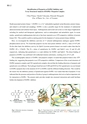
BG-2Identification of Purpurin as DAPK1 Inhibitor andX-ray Structural Analysis of DAPK1?Purpurin Complex○Peter Wijaya, Takeshi Yokoyama, Mineyuki Mizuguchi(Fac. of Pharm. Sci., Univ. of Toyama)Death-associated protein kinase 1 (DAPK1) is a Ca 2+ /calmodulin-regulated serine/threonine protein kinase,and related to cell death and autophagy. DAPK1 is also a possible target for the treatment of endometrialadenocarcinomas and ischemic brain injury. Anthraquinone and its derivatives have a wide range of applicationincluding for medical and therapeutic applications, such as antineoplastic and antiarthritic agent. In recentstudies, quinalizarin (anthraquinone derivative) has been reported as an ATP-competitive inhibitor of proteinkinase CK2. This could be useful as starting point for development of various protein kinase inhibitor.Here, we investigated the inhibitory activities of 17 selected anthraquinone analogues against DAPK1phosphorylation activity. We found that purpurin was the most potent inhibitor with IC 50 value of 0.85μM.On the other hand, the inhibitory activity for EphA3 (tyrosine protein kinase) was much weaker than that forDAPK1 (IC 50 >120μM). The IC 50 values of quinalizarin for DAPK1 and EphA3 were 10 and 44μM,respectively, indicating that quinalizarin is a weak inhibitor for DAPK1 and EphA3. The direct binding ofpurpurin to DAPK1 was confirmed with isothermal titration calorimetry, resulting in K d of 0.21μM.The crystallographic analysis of DAPK1 and purpurin complex revealed that purpurin binds to the ATPbindingsite, suggesting that purpurin is an ATP-competitive inhibitor. Comparison of the crystal structures ofDAPK1-purpurin complex and CK2-quinalizarin complex showed that the binding directions of purpurin andquinalizarin were different. The hydrogen bond between 8-OH and H160 in CK2-quinalizarin was not formedin DAPK1-purpurin due to the mutation (E143 in DAPK1) and the different binding direction. This would bethe reason why the inhibitory potency of quinalizarin against DAPK1 was weaker than that of purpurin. It wasindicated that the presence and position of hydroxyl group in anthraquinones derivatives had an important rolefor interaction to DAPK1. The present study provides insight into structural interaction and could facilitatefurther development of DAPK1 inhibitor.purpurinIC 50 = 0.85μM (DAPK1)>120μM (EphA3)quinalizarinIC 50 = 10μM (DAPK1)44μM (EphA3)Fig. 1. Molecular structures of purpurinand quinalizarin.Fig. 2. Superimposition of DAPK1-purpurin complex ( ) andCK2-quinalizarin complex ( PDB code: 3FL5, ). Numbers representthe positions of hydroxyl group.